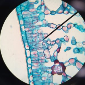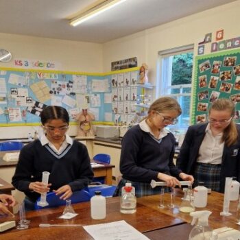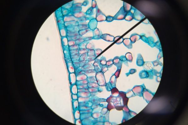
Practical Science lessons are already in full swing at Braeside. In a Year 10 lesson, Amaarah took a photo of her light microscope image of a cross section of a leaf, magnification x 100.
The pointer identifies the Palisade mesophyll layer and the spongy mesophyll is clear underneath with plenty of air spaces for the efficient diffusion of gases into and out of the leaf.
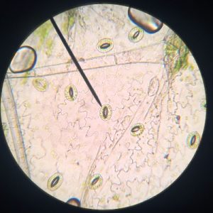
Alex and Mackensie managed to create a light microscope image of their lower epidermal leaf tissue which they carefully peeled from a Geranium leaf using forceps. They did really well to get the detail of the lower epidermal cells. You can clearly see about 9 stomata openings surrounded by their guard cells. The stomata are open to allow for gas exchange.
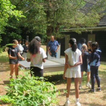
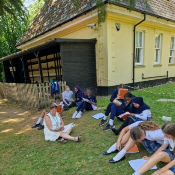
Pupils found the lesson very interesting, and the practical approached helped with furthering their understanding of the topic.